Guide to Nudibranch Body Parts
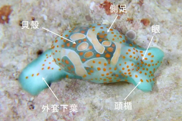
To identify nudibranchs accurately, it's essential to understand their body parts. This guide explains the key anatomical features used in nudibranch identification.
Rhinophores (触角)
Rhinophores are the pair of sensory organs on top of the nudibranch's head. They detect chemical signals in the water and are crucial for finding food and mates. Their shape — smooth, lamellate, or perfoliate — is an important identification feature.
Secondary Gills (二次鰓)
The feathery structures on the back of dorid nudibranchs are secondary gills (branchial plume). They're used for respiration and surround the anus. The number and arrangement of gill branches varies by species.
Mantle (外套膜)
The mantle is the fleshy covering on the dorsal side. In many dorids, it extends beyond the foot as a "mantle skirt." The texture, color patterns, and border of the mantle are key identification features.
Oral Tentacles (口触手)
Located near the mouth, oral tentacles help the nudibranch sense food at close range. Their shape and size differ significantly between families.
Cerata (突起)
Aeolid nudibranchs have finger-like projections called cerata instead of gills. These contain branches of the digestive gland, and in some species, stored nematocysts (stinging cells) from their cnidarian prey.
Foot (足)
The muscular underside used for locomotion. The foot's shape, width, and coloration can help with identification, though it's often not visible in photographs.
Enjoyed this post? You can tip the author directly —
Tip this post