Are nudibranch colors really "structural"? — Multiple pigment, kleptoplast, and photonic pathways in the primary literature
In April 2026, several Japanese popular-science sites ran the headline "Nudibranch colors turned out to be structural colors." The underlying paper is Humphrey et al. (2026) in PNAS, which shows that the body color of certain nudibranchs comes from photonic structures made of guanine nanocrystals.
Two things are misleading in those popular-science framings. First, the paper studied six species — four chromodorids and two aeolidiids — not "nudibranchs" as a whole. Second, "previously thought to be pigment-based" is only partly true. Many nudibranchs still get their color from pigments — often sequestered from prey — and the green sacoglossans get their color from stolen chloroplasts (kleptoplasty), not pigments and not photonic crystals.
This post walks through what Humphrey et al. (2026) actually demonstrated, and what the three main independent pathways of nudibranch coloration look like in the primary literature.
What Humphrey et al. (2026) actually showed
The full title is "Nudibranch color diversity shares a common physical basis in guanine photonic structure 'pixels'." The study is a collaboration between the Max Planck Institute of Colloids and Interfaces in Potsdam and the University of Cambridge, with Samuel Humphrey as first author and Silvia Vignolini as senior author.
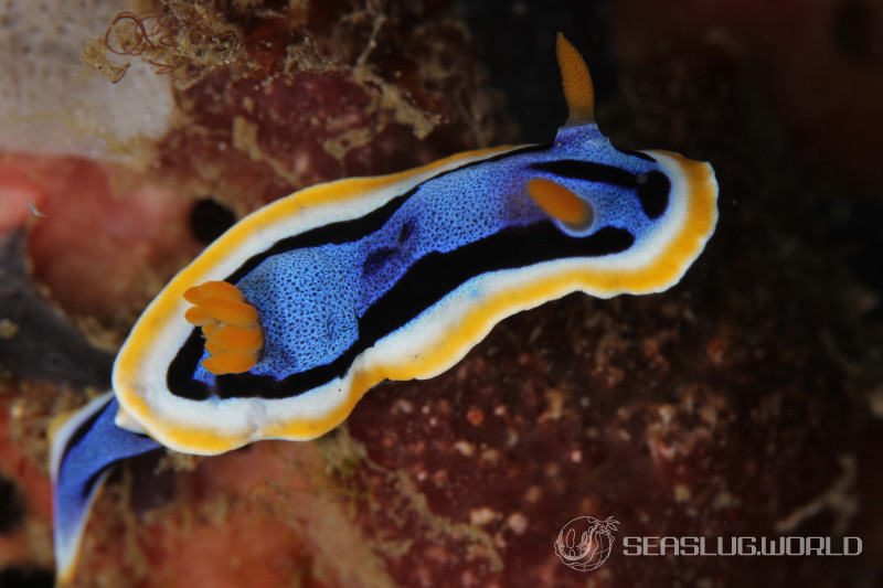
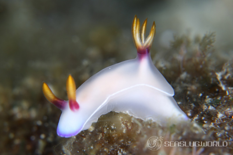
They examined six species: Chromodoris annae, Chromodoris willani, Hypselodoris bullockii and Hypselodoris tryoni (Chromodorididae), plus Spurilla neapolitana and Berghia stephanieae (Aeolidiidae). The cross-clade selection matters because one of the paper's claims is that this color mechanism is a "common physical basis" shared across very different lineages (Doridida vs. Cladobranchia). The methodological centerpiece is cryogenic focused ion beam (cryo-FIB) SEM tomography, which let them reconstruct the three-dimensional arrangement of guanine nanoplatelets within the skin.
The novel claim is not that structural color exists in nudibranchs — structural color in molluscs has been recognized for decades — but that guanine multilayers in these species are organized into "pixel"-like domains. Within each pixel the nanoplatelets are slightly disordered in orientation, which damps angle dependence and produces a matte structural color rather than an iridescent one. The paper argues this matters functionally because aposematic warning coloration works better when the color looks the same from every angle.
Three independent pathways for nudibranch coloration
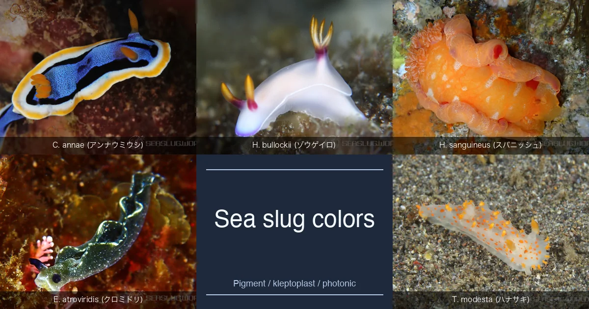
"Nudibranchs" in the broad sense include roughly 3000 known species. The mechanisms behind their colors are not unified — at minimum there are three distinct routes.
Pathway 1: Dietary pigments (the majority of species)
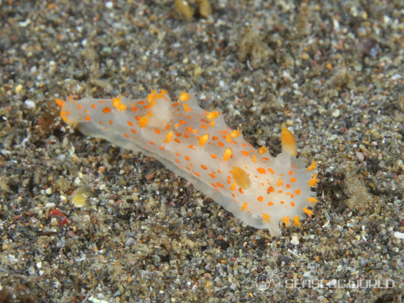
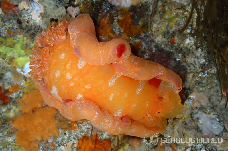
Dorid nudibranchs, including chromodorids, are well known to sequester secondary metabolites from sponge prey for chemical defense; Cimino & Ghiselin (2009), "Chemical defense and the evolution of opisthobranch gastropods" (Proc. Cal. Acad. Sci. 60: 175-422), is a standard review of opisthobranch chemical ecology. Pigments often travel through the same route. The orange of Triopha catalinae (formerly T. carpenteri) is triophaxanthin, an acetylenic apocarotenoid considered to be of dietary origin. The red pigment hurghadin in the Red Sea Hexabranchus sanguineus is likewise a sequestered carotenoid. These colors are pigment-based, not structural.
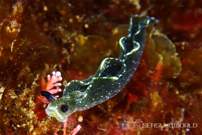
Pathway 2: Kleptoplasty (Sacoglossan greens)
The sacoglossan sea slugs retain chloroplasts from algal prey inside their digestive cells. Elysia chlorotica keeps chloroplasts of the yellow-green alga Vaucheria litorea functional for months, evolving photosynthetic oxygen without further feeding. Rumpho et al. (2008) in PNAS reported expression of algal-derived psbO in the slug, but subsequent genomic work (e.g. Bhattacharya et al. 2013) strongly disputed the horizontal gene transfer interpretation, and the debate continued. In 2021 Maeda et al. (eLife 10: e60176) showed that Plakobranchus ocellatus achieves functional kleptoplasty without HGT. Either way, the green of these sea slugs is literally the chloroplast itself — not a pigment of the slug, not a photonic crystal.
Pathway 3: Guanine structural color (Humphrey et al. 2026)
This is where the new PNAS paper sits. Padula et al. (2016) had earlier shown, in the Felimida clenchi complex, that color patterns are not strictly phylogenetically informative and may represent mimicry circles — suggesting that similar color "solutions" can be reached convergently. Humphrey et al. now provide the physical basis for at least one of those solutions: pixel-organized guanine multilayers giving angular-independent matte structural color.
What is misleading in the popular framing
- "Nudibranch color is structural color." Overgeneralization. The study is six species (four chromodorids and two aeolidiids), focused on blues. Sacoglossan greens, dorid oranges, and many reds in other groups are not structural.
- "Colors were previously thought to be pigments." Partly wrong. Pigment-based color is still the dominant mechanism across the many lineages, and that chemistry has been studied for decades.
- "Nanocrystals make the colors." Correct, for the species the paper actually examined.
Why angular independence matters
There are two broad regimes of structural color:
- Iridescence, where the color shifts strongly with viewing angle (morpho butterflies, peacock feathers, CD surfaces). This requires long-range ordered lattices. In plants the blue fruit of Pollia condensata (Vignolini et al. 2012) is a striking "pixelated" iridescent example.
- Matte structural color, where deliberate disorder in the same nanostructures erases angle dependence. The classic case is the non-iridescent blue of Blue Jay and Cotinga feathers, produced by quasi-ordered keratin nanostructures inside the feather barb (Saranathan et al. 2012, J. R. Soc. Interface).
For aposematism, matte is preferable: a predator learning "blue band = bad" can only generalize if the band looks blue from every direction. Humphrey et al.'s contribution is the 3-D structural recipe for matte color using guanine — a specific orientational-disorder design at the pixel scale.
On seaslug.world
Our Colors hub lets you browse species by primary color, but this is a visual filter, not a mechanism filter. A "blue" Chromodoris and a "blue" translucent deep-sea aeolid are blue for completely different reasons. Where possible, the species description mentions the likely mechanism (kleptoplasty for sacoglossans, guanine structural color for chromodorid blues). Photographers of the group might enjoy thinking about which mechanism produces the color they captured.
Related links
Enjoyed this post? You can tip the author directly —
Tip this post